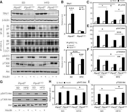
FIG. 4.
Protected insulin signaling in Ptpn6H-KO mice. A–F: Western blot analysis of liver lysates from Ptpn6f/f and Ptpn6H-KO mice fed the SD or HFD for 8 week, fasted 6 h, and followed by tail vein administration of either saline or insulin (n = 12–13 per genotype and diet group; ^P < 0.05 and ^^^P < 0.005 diet effect, *P < 0.05, **P < 0.01, and ***P < 0.005 genotype difference). Dotted line on blot borders shows noncontiguous sections of the same gel. Shp1, pS473, pT308, and total Akt were detected directly using their respective antibodies, with β-actin as the loading control. IR and CC1-L were immunoprecipitated before being immunoblotted for pY, total IR, and CC1. G–I: Phosphorylation of Akt (pS473 and pT308) and total Akt in purified hepatocytes isolated from Ptpn6f/f and Ptpn6H-KO mice (8 weeks SD or HFD) were detected by immunoblotting with respective antibodies. Purified hepatocytes were cultured for 16 h, serum-deprived for 3 h, and treated with either PBS or insulin for 15 min before cell lysis (n = 3 per genotype and diet group; ^P < 0.05 diet effect; *P < 0.05 genotype difference).

