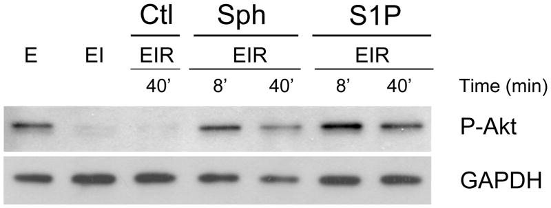
Figure 2. Effect of Postconditioning with S1P and Sphingosine on the Phosphorylation of Akt.
Ex vivo hearts were either equilibrated for 20’ (E), equilibrated followed by 40 min of ischemia (EI), or equilibrated followed by 40 min of ischemia followed by reperfusion (EIR). For the vehicle control (Ctl), reperfusion was for 40 min with medium containing vehicle only (Ctl EIR-40’). For postconditioning, after 40 min of global ischemia (EI), hearts were reperfused with medium containing either 0.4 μM S1P or 0.4 μM sphingosine for either 8 min or 40 min (EIR-8’and EIR-40’). After 40 min of reperfusion, the hearts were collected, homogenized and separated into cytosolic and particulate fractions. The Figure shows the western analysis for the cytosolic fraction using an antibody to phospho-AKT (ser473). Each lane was loaded with 10 μg of protein. The lanes were loaded as follows, lane 1- equilibrated; lane 2-equilibrated plus ischemia; lane 3-equilibrated plus ischemia plus vehicle reperfusion for 40’: lane 4- 8’ of reperfusion in the presence of sphingosine, lane 5– 40’ of reperfusion in the presence of sphingosine,, lane 6– 8’ of reperfusion in the presence of S1P, lane 7– 40’ of reperfusion in the presence of S1P.

