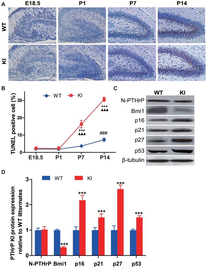
Figure 4. PTHrP NLS and C terminus deficiency increases neural cell apoptosis and up-regulate expression levels of CDKIs in the hippocampus.
(A) Representative micrographs of sections from the hippocampus of brains from E18.5, P1, P7 and P14 WT and KI mice stained for apoptotic cells using the TUNEL technique (brown, magnification, ×400). (B) The percentage of apoptotic cells in hippocampus was quantified by computer-assisted image analysis. (C) Western blot analysis of hippocampus protein extracts for N-terminal PTHrP (N-PTHrP), Bmi1, p16, p21, p27 and p53. β-tubulin was used as loading control. (D) N-PTHrP, Bmi1, p16, p21, p27 and p53 protein levels relative to β-tubulin protein level were assessed by densitometric analysis and expressed relative to levels of WT mice. Each value is the mean±SEM of determinations in 5 mice of each group. ***, P<0.001 in Pthrp KI mice relative to wild-type littermates. ###, P<0.001 at the time point relative to the prior observed time point in WT mice. ▴▴▴, P<0.001 at the time point relative to the prior observed time point in Pthrp KI mice.

