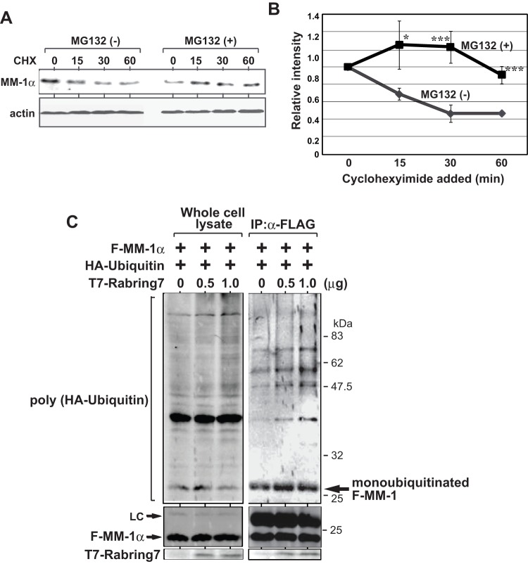
Figure 2. Mono-ubiquitination of MM-1 by Rabring7.
A. HeLa cells were cultured in the presence or absence of cycloheximide. At the times indicated, the expression levels of MM-1α and actin in cells were examined by Western blotting with respective antibodies. B. The intensity of bands corresponding to MM-1α and to actin in Fig. 2A was quantified, and relative intensity of MM-1α to that of actin is shown. Values are means ± S.D. n = 3 experiments. Significance: *p<0.05 and ***p<0.001. C. HEK293T cells were transfected with FLAG-MM-1α together with HA-ubiquitin and with various concentrations of T7-Rabring7. Forty-eight hrs after transfection, proteins prepared from transfected cells were immunoprecipitated with an anti-FLAG antibody, and the precipitates were analyzed by Western blotting with anti-HA, anti-FLAG and anti-T7 antibodies (right panel). A part of the proteins prepared from transfected cells was also analyzed (left panel).

