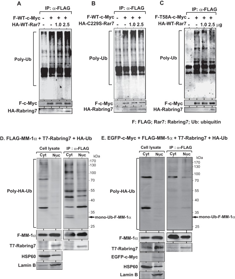
Figure 6. Stimulation of poly-ubiquitination of c-Myc by Rabring7.
A and B. HEK293T cells were co-transfected with FLAG-wild-type-c-Myc together with two doses of HA-wild-type Rabring7 (A) or HA-C229S-Rabring7 (B). Forty-four hrs after transfection, 25 μM MG132 was added to the culture medium, and the cells were cultured for an additional 4 hrs. Proteins in cells were then immunoprecipitated with an anti-FLAG antibody, and the precipitates were analyzed by Western blotting with an anti-multi-ubiquitin antibody. C. HEK293T cells were co-transfected with FLAG-T58A-c-Myc together with two doses of HA-wild-type Rabring7 and subjected to ubiquitination assays as described in the legends of Figures 6A and 6B. D. HEK293T cells were co-transfected with FLAG-MM-1α, T7-Rabring7 and HA-ubiquitin. Forty-eight hrs after transfection, total cell lysates were prepared and the cytoplasm and nucleus were fractionated as described in Experimental procedures. Proteins extracted from them were immunoprecipitated with an anti-FLAG antibody and analyzed by Western blotting with anti-HA, anti-FLAG and anti-T7 antibodies. Fractions of the cytoplasm and nucleus were also blotted with anti-HSP60 (SC-6216, Santa Cruz biotechnology, Santa Cruz, CA) and anti-Lamin B (SC-13115, Santa Cruz biotechnology) antibodies. E. HEK293T cells were co-transfected with EGFP-c-Myc, FLAG-MM-1α, T7-Rabring7 and HA-ubiquitin. Proteins were analyzed as described in the legend of Figure 6D.

