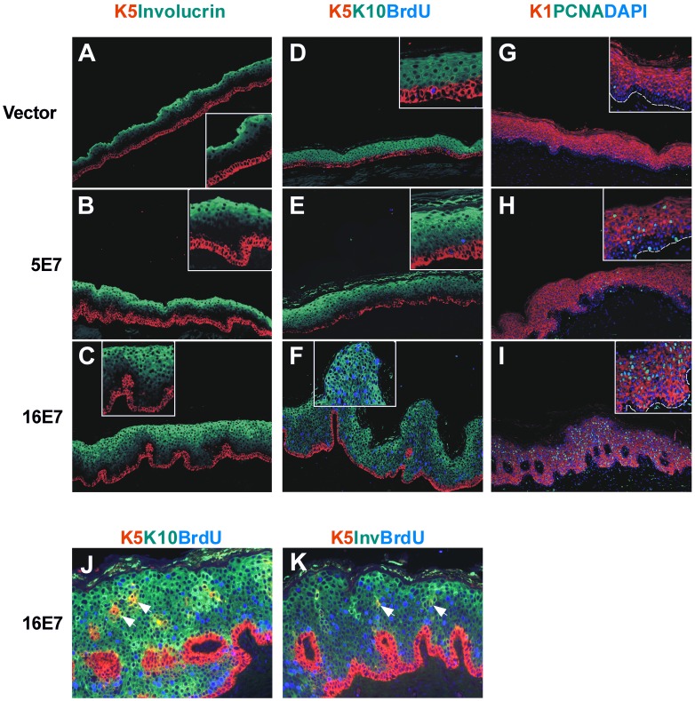
Figure 3. Epidermal differentiation and proliferation markers in E7-grafts.
Sections of paraffin-embedded grafts were processed for immunofluorescence staining with antibodies to epithelial differentiation markers (basal cytokeratin 5, early suprabasal cytokeratin 1 and 10, and late suprabasal involucrin) and proliferation markers (BrdU, PCNA). As in normal skin, cytokeratin 5 (K5) is expressed in the basal layer, cytokeratin 10 (K10) and 1 (K1) in early suprabasal cells, and involucrin (Inv) in late differentiated cells in control vector samples (A, D and G). A similar phenotype was observed in the HPV5 E7 samples (B, E and H). An expansion of involucrin positive cells downwards was observed in HPV16 E7-transplants (C and K). BrdU and PCNA, normally present in some basal cells (D and G), are ectopically present in suprabasal cells of HPV16 E7 samples (F and I). Focal areas of suprabasal PCNA positive cells occur in HPV5 E7 samples (H). Importantly, suprabasal K5/K10 positive (J) or K5/involucrin positive cells (K) were observed in 16E7-grafts. Where indicated, DAPI staining was used to visualize cellular nuclei. K5Inv: staining with both K5 and involucrin antibodies; K5K10BrdU: staining with K5, K10 and BrdU antibodies; K1PCNADAPI: staining with K1, PCNA and DAPI. Dashed line indicates the location of the basal membrane.

