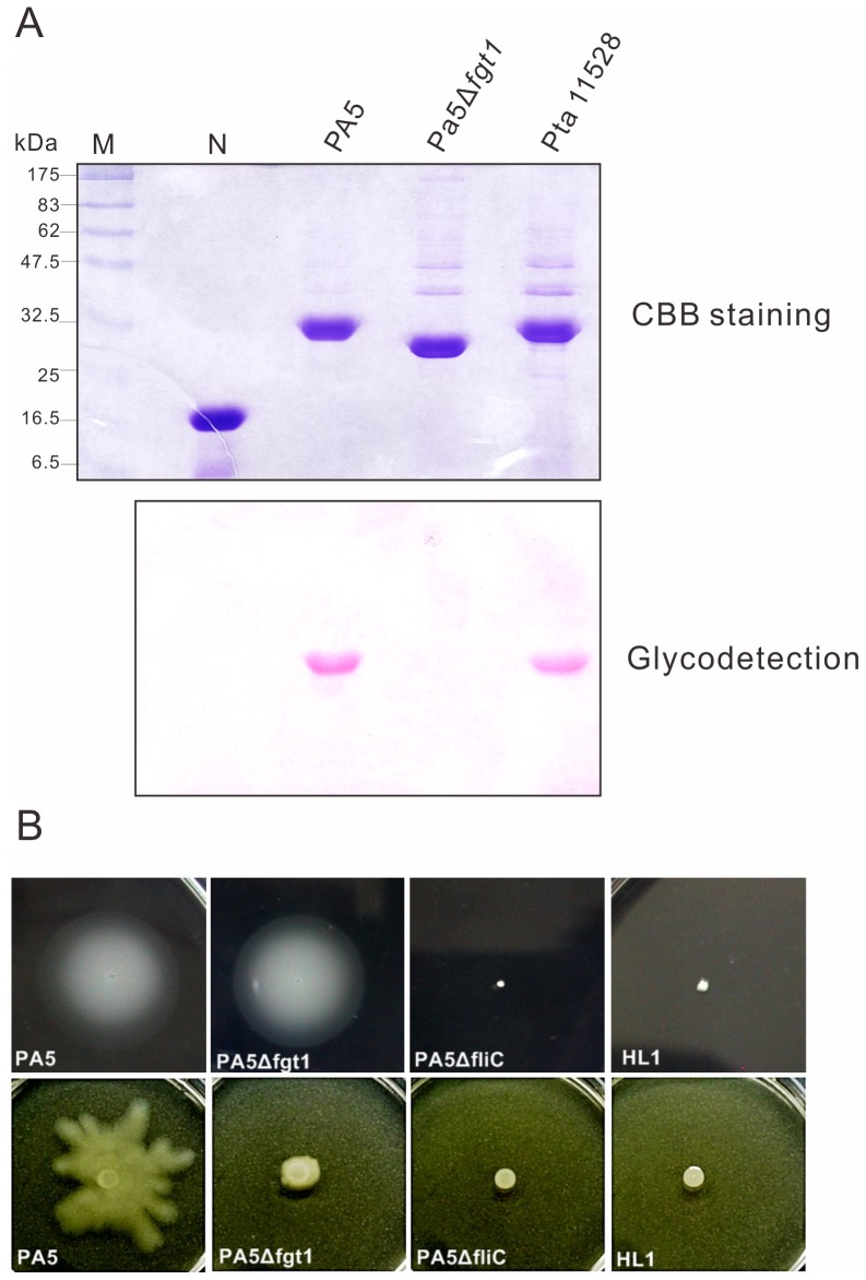
Figure 6. Flagellin glycosylation and motility of wild type (PA5) and the fgt1 mutant (PA5Δfgt1).
(A) Staining on glycosylated flagellin. Purified proteins separated with SDS-12%-PAGE were stained by Coomassie brilliant blue (CBB) (upper panel) and by using a GelCode glycoprotein staining kit (Pierce, Rockford, III) (lower panel). Soybean trypsin inhibitor was served as a negative control (N) and purified flagellin from P. s. pv. tabaci (Pta) was a positive control. (B) Motility assay. For swimming assays (upper), bacterial strains were incubated for 2 days at 23°C on 0.3% soft agar of hrpMM plate. For swarming assays (bottom), bacterial strains were inoculated on 0.5% SWM agar plate and were observed after 24 h at 28°C.

