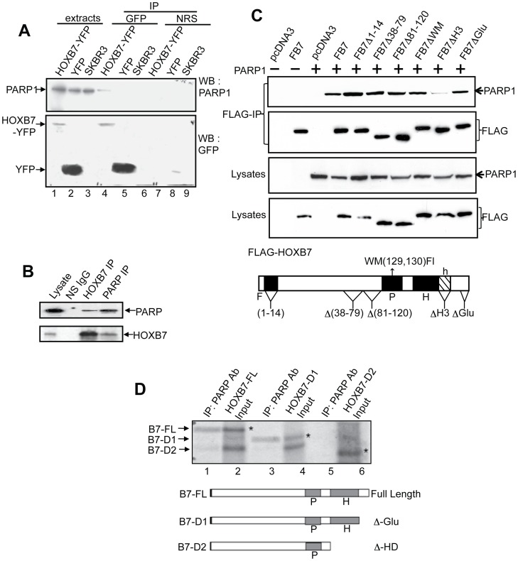
Figure 1. HOXB7 interacts with PARP-1.
A. Co-immunoprecipitation of PARP-1 with HOXB7-YFP in SKBR3 cells. SKBR3 cells were stably transfected with HOXB7-YFP (lanes 1, 4, and 7) or YFP alone as a vector control (lanes 2, 5, and 8) prior to immunoprecipitation with GFP antibodies and subsequent Western blotting of precipitated proteins. Parental SKBR3 cells, which lack detectable HOXB7, were used as controls as well (lanes 3, 6, and 9). Lanes 1 to 3, protein levels in 100µg of total cell extract (5% of input); lanes 4 to 6, proteins that precipitated with HOXB7-YFP or controls that did not express HOXB7 (SKBR3-YFP and parental cells). Normal rabbit serum (NRS) was used to control for specificity (lanes 7–9). B. Endogenous interaction between HOXB7 and PARP-1. Extracts of MCF-7 cells were co-immunoprecipitated with antibodies to PARP-1, HOXB7 or p53 as a nonspecific IgG (NS IgG). Subsequent immunoblotting was done with the antibodies indicated. C. Flag-tagged HOXB7 or constructs in which select regions were deleted or mutated, were transiently transfected into CHO cells together with a PARP expression construct (PARPpCR3.1, where indicated, empty plasmid was used as control) to determine if a specific region of HOXB7 mediated its interaction with PARP. Co-immunoprecipitation with FLAG antibodies (top panel) was performed followed by immunoblot with PARP antibodies. The lower panel shows protein expression of all transfected plasmids. Structure of FLAG-HOXB7 showing locations of point mutations and deletions is shown below. F: Flag tagged HOXB7, P: pentapeptide, H: homeodomain, h: helix 3 of the homeodomain. D. Full length HOXB7 or deletion constructs B7-D1 or B7-D2 were transcribed and translated in vitro in the presence of 35S-methionine prior to mixing with in vitro transcribed and translated PARP-1. Immunoprecipitation was performed with PARP-1 monoclonal antibodies (lanes 1, 3 and 5). Complexes were resolved by SDS-PAGE and subjected to autoradiography for 24 hours. Lanes 2, 4 and 6 are input (20%) from the TNT reactions. Deletions D1 and D2 of full length HOXB7 are shown in Figure 2. Asterisks point to the specific bands for HOXB7 full-length or deletion proteins. P: pentapeptide, H: homeodomain.

