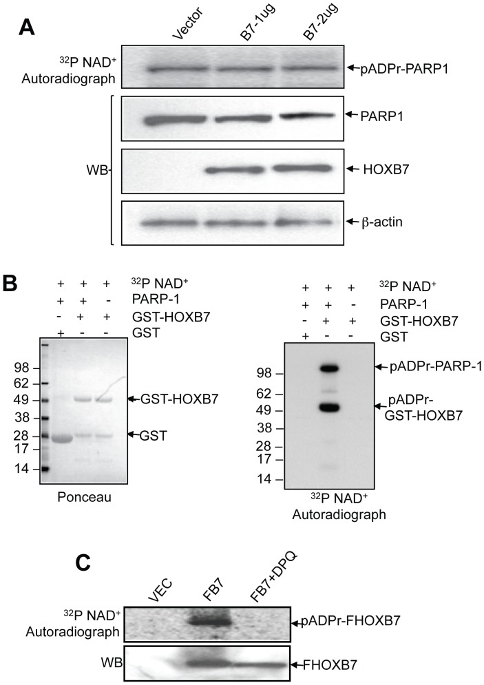
Figure 3. HOXB7 is the substrate of PARP-1 and is poly(ADP ribosyl)ated by PARP-1.
A. Different amounts of Flag tagged HOXB7 plasmids were transfected into SKBR3 cells, cells were harvested and permeabilized by 0.01% digitonin in PARP-1 activity reaction buffer. PARP-1 auto-modification was visualized by autoradiograph after the cell lysates were separated by SDS-PAGE (top panel). Immunoblot with anti-Flag, PARP-1 and β-actin antibodies were performed after autoradiography (bottom panels). B. GST and GST-HOXB7 fusion proteins were incubated with or without purified PARP-1 protein or 32P NAD+. After 30 minutes incubation, free 32P NAD+ and unbound PARP-1 proteins were washed off with reaction buffer. Proteins on glutathione sepharose beads were then separated by SDS-PAGE and transferred to PVDF membrane and stained with Ponceau (left panel). The poly(ADP ribosyl)ated proteins were visualized by autoradiography (right panel). C. Vector control and Flag-tagged HOXB7 plasmids were transfected into SKBR3 cells. Cells were harvested and incubated with PARP-1 activity assay buffer including 32P NAD+ or PARP-1 inhibitor (20 µM DPQ). Cell lysates were immunoprecipitated with anti-Flag antibody and separated by SDS-PAGE followed by autoradiography (top panel) and immunoblotting with anti-Flag antibodies (bottom panel).

