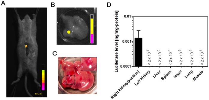
Figure 2. In vivo transfection of naked pDNA by tissue suction.
A) In vivo imaging of luciferase activity in a mouse liver that was suctioned once by the type I device just after intravenous injection of pCMV-Luc. B) Ex vivo imaging of luciferase activity in the liver suctioned by the type I device. C) Bright field image of (B). D) Luciferase levels of various tissues. The right kidney in mice was suctioned once by the type III device. Each value represents means + SD (n = 4). All mice were alive at the end of the experiment.

