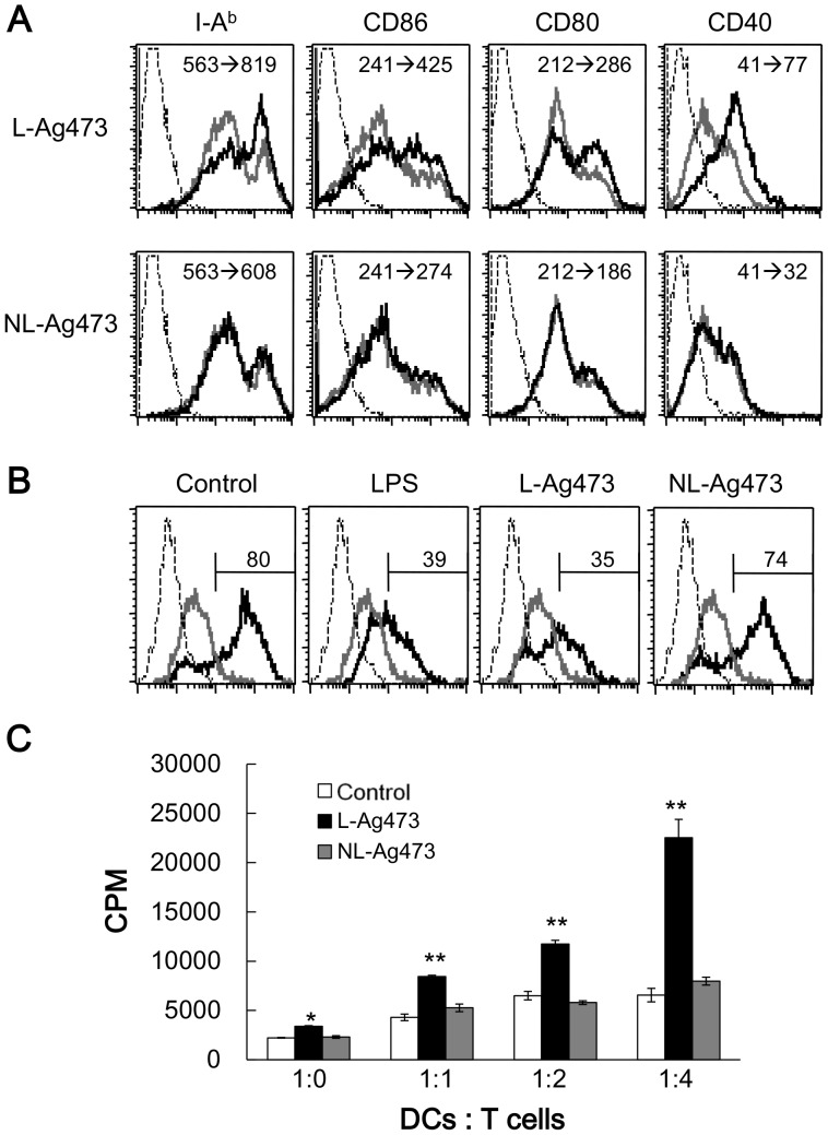
Figure 6. L-Ag473 promotes BMDC maturation and DC-induced T cell activation.
(A) BMDCs were treated with L-Ag473, NL-Ag473 (100 ng/ml) (black line), or untreated (gray line) for 16 hours, and then stained with anti-I-Ab, CD86, CD80, and CD40 mAbs. Dotted line represents staining with an isotype-matched control Ab. The change of mean fluorescence intensity (MFI) from no treatment to Ag473 treatment is shown on each picture. (B) BMDCs were treated with LPS (20 ng/ml), L-Ag473, NL-Ag473 (100 ng/ml), or untreated for 16 hours. The ability of endocytosis of DCs was determined by the uptake of dextran-FITC at 4°C (gray line) or 37°C (black line). Dotted line represents untreated DCs without dextran-FITC. The percentages of dextran-FITC+CD11c+ cells were shown above the regional markers. All data are representative of two to four independent experiments. (C) CD4+ OT-II T cells were isolated and co-cultured with L-Ag473 or NL-Ag473 (100 ng/ml)-activated DCs pulsed with OVA peptide (2 µg/ml) at indicated ratio of DC: T cell for 72 hours. T cell proliferation was determined by [3H]thymidine incorporation. Data are shown as mean + SD from triplicate DC cultures; *p<0.05; **p<0.01 (Student’s t-test), comparing L-Ag473-treated to non-treated cells.

