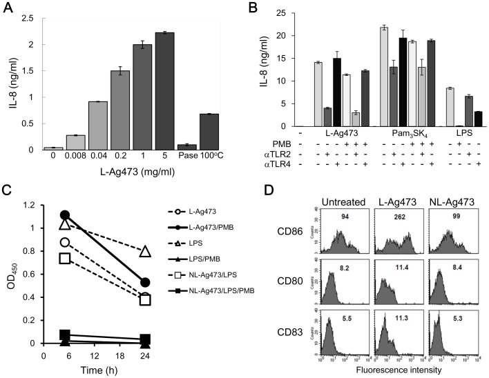
Figure 9. The effect of L-Ag473 on human cells.
(A) THP-1 cells (2×10⁵/well) were incubated with the indicated amounts of L-Ag473, proteinase K-treated (PNaseK), heated (100°C) L-Ag473 (5 µg/ml). (B) THP-1 cells (2×10⁵/well) were incubated with L-Ag473 (1.5 µg/ml), Pam3SK4 (100 ng/ml) (InvitroGen, Cat No. tlr1-pms) or LPS (100 ng/ml) (Sigma, Cat No. L4391) alone (−PMB) or pretreated with PMB (+PMB) for 18 hours. For antibody blocking experiments, THP-1 cells were preincubated with the indicated antibody for 30 min before stimulation. IL-8 was undetectable in the untreated cells (−). (C) PBMCs (1×10⁶/well) were incubated with the indicated reagent. TNF-α in the 5-h and 24-hours culture supernatants were determined by ELISA. The result shown is the reading values with the untreated cells as the blank. (D) MoDCs were untreated as control, or incubated with L-Ag473 or NL-Ag473 mutant (100 ng/ml) for 16 hours, and then stained with PE-anti-CD86, FITC-anti-CD80, and PE-anti-CD83 mAbs. The MFI is shown on each picture. The data are representative of three independent experiments.

