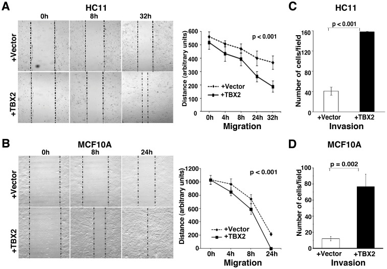
Figure 2. TBX2 promotes migration and invasion of mammary epithelial cells.
(A, B) In vitro ‘scratch’ assays (see Methods) monitoring the migration of (A) murine HC11 and (B) human MCF10A cells stably expressing pCDNA3 vector (+vector) or pCDNA3-TBX2 (+TBX2) over a period of 24–32 hours (h). Representative bright field images of cells (10x magnification) are shown in the left panel. Right panel: statistical evaluation of the distance between the two borders (dotted lines; left panels) at different time points after ‘scratch’ (n = 3; ANOVA test). (C, D) Transwell matrigel invasion assays show a significantly increased ability of TBX2-expressing HC11 (C) and MCF10A cells (D) to invade through a matrigel layer (n = 3, Student’s t-test). The mean ± S.D. is shown. P values are indicated.

