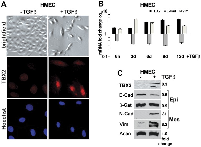
Figure 3. Endogenous TBX2 is induced during TGFß-mediated EMT of primary human mammary epithelial cells (HMEC).
(A) Bright field (40x magnification) and immunofluorescence images (63X magnification) of primary HMEC treated with 5 ng/ml TGFß1 (+TGFß) for 12 days as compared to untreated control cells (-TGFß). TGFß induces EMT-like morphological changes and nuclear expression of TBX2 (red). Nuclei were stained with Hoechst 33258 (blue). (B) qPCR analysis shows a time course analysis of TBX2 mRNA induction in TGFß–treated HMEC in comparison to changes in epithelial E-cadherin (E-cad) and mesenchymal Vimentin (Vim) expression. Values were normalized to GAPDH mRNA and represent fold changes as compared to control untreated HMEC at the indicated time points. Error bars represent SEM of each sample in triplicates. (C) Western blot analysis of TBX2 and EMT marker expression in untreated HMEC (−) and in HMEC treated with TGFß for 12 days. E-cad = E-cadherin; ß-cat = ß-catenin; Vim = Vimentin; N-cad = N-cadherin; Epi = epithelial; Mes = mesenchymal markers. Actin = ß-actin was used as loading control. Densitometric quantification of fold changes in protein levels normalized to Actin values is shown.

