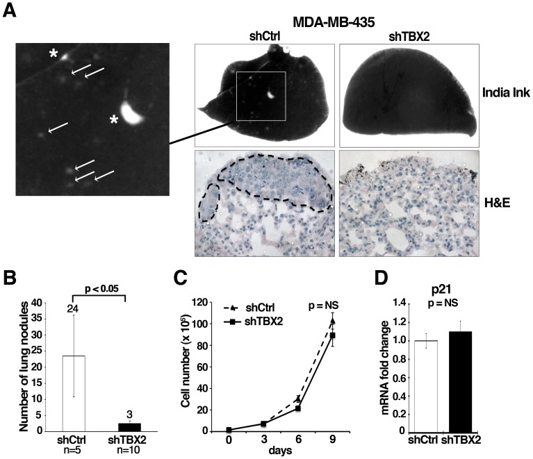
Figure 7. Knockdown of TBX2 reduces pulmonary metastasis of human MDA-MB-435 breast carcinoma cells.
(A) Representative images of lungs harvested from athymic nu/nu Nude mice forty days after tail vein injection with MDA-MB-435 tumor cell clones expressing either control non-target shRNA (shCtrl) or TBX2-specific shRNA (shTBX2). Top panel: India ink staining of lungs shows the absence of surface lung metastases in mice injected with shTBX2-expressing MDA-MBA-435 tumor cells (Magnification 7x). Only the control group produced macroscopic lung nodules (asterixes) and an elevated number of micrometastases (white arrows). Bottom panel: H&E stained paraffin-sections of representative lungs from each study group (Magnification: 40X). Dotted lines highlight lung metastases in the control group. (B) Quantification of total lung metastasis burden in the same sets of mice as in (A). Average numbers of lung surface metastases are shown; white column = mean of 5 control mice analyzed: black column = mean of 10 mice injected with two MDA-MB435-shTBX2 tumor cell clones. Data represent the mean ± S.D. (n≥5; Student t-test). (C) Inhibition of TBX2 does not significantly affect cell proliferation of MDA-MB-435 tumor cells. Equal numbers of control non-target shRNA and shTBX2-expressing cells were grown under sub-confluent conditions and counted every 3 days over a 9-day period. Error bars represent the mean ± S.D. (n = 3; Student t-test). (D) qPCR showing that stable knockdown of TBX2 does not significantly alter p21 mRNA expression levels in MDA-MB-435 tumor cells. Values were normalized to GAPDH and fold changes compared to the shRNA control group are shown. Error bars represent the mean ± SEM (n = 3; Student t-test). NS = not significant.

