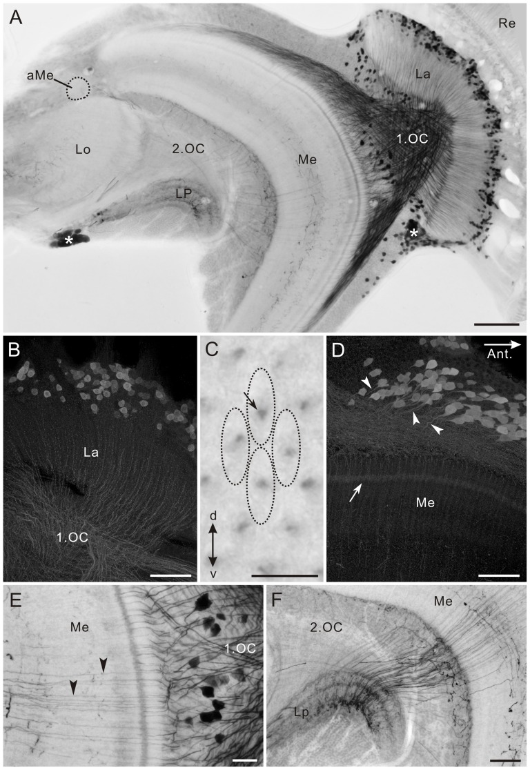
Figure 6. Tyramine immunoreactivity in the optic lobe of Papilio xuthus.
A: Horizontal section. The accessory medulla (aMe) is free of labeling. Asterisks indicate remnants of larval stemmata. B: Horizontal section of the lamina (La). Large monopolar cells (LMCs) exhibiting tyramine immunoreactivity project thin axons through the lamina into the medulla. C: Lamina cross section with axon profiles of tyramine-positive LMCs (arrow). Each cartridge (dotted ovals) contains one tyramine-positive axon. d, dorsal; v, ventral. D: Horizontal section of the anterior medulla. Numerous tyramine-positive neurons probably correspond to medulla amacrine cells. Their primary neurites enter the medulla (arrowheads). Layer 2 exhibits moderate staining (arrow, also see Fig. 7D–F). E,F: Horizontal sections. E: Thin fibers from cell bodies in the first optic chiasma (1.OC) pass through the medulla (arrowheads) and terminate in both outer and inner layers of the lobula plate (LP, F). 2.OC, second optic chiasma; Lo, lobula; Re, retina. Scale bar = 100 µm in A; 50 µm in B,D,F; 10 µm in C; 20 µm in E.

