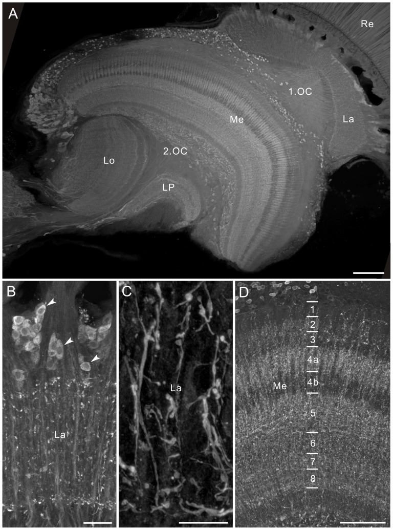
Figure 9. GABA immunofluorescence in the optic lobe of Papilio xuthus, horizontal sections.
A: GABA-positive cell bodies are located in the lamina cell body layer, at the anterior rim of the medulla (Me), along the distal surface of the medulla, and between the medulla and lobula plate (LP). B: Longitudinal section of the lamina (La). Some LMCs are GABA-positive (arrowheads). C: Enlarged image of the lamina synaptic layer, showing GABA-positive longitudinally-oriented fibers with small collaterals and beaded terminals. D: All layers of the medulla are innervated by GABA-positive fibers. 1.OC, first optic chiasma; 2.OC, second optic chiasma; Lo, lobula; Re, retina. Scale bar = 100 µm in A; 20 µm in B; 10 µm in C; 50 µm in D.

