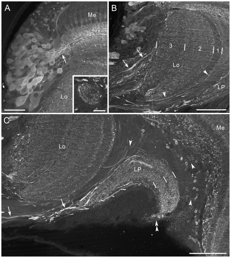
Figure 11. GABA immunofluorescence in the medulla, lobula and lobula plate.
Horizontal sections. A: A cluster of GABA-positive cell bodies at the anterior rim of the medulla (Me) projects primary neurites into layer 5 (arrow). Inset shows a single optical section containing the accessory medulla, innervated by GABA-positive fibers. B: The lobula (Lo) is innervated by GABA-positive thick processes from the central brain (arrows; anterior to the top, medial to the left). The processes bear numerous fine fibers, distributed throughout the lobula. Arrowheads indicate thin fibers running along the distal surface of the lobula. C: The lobula plate (LP) is invaded by thick processes (arrows), and both layers are densely innervated by fine fibers. Arrowheads indicate thin fibers linking the medulla, lobula and lobula plate. GABA-positive cell bodies are located between the medulla and lobula plate, and at the posterior margin of the lobula plate (double arrowhead). Scale bar = 50 µm in A; 20 µm in inset of A; 100 µm in B,C.

