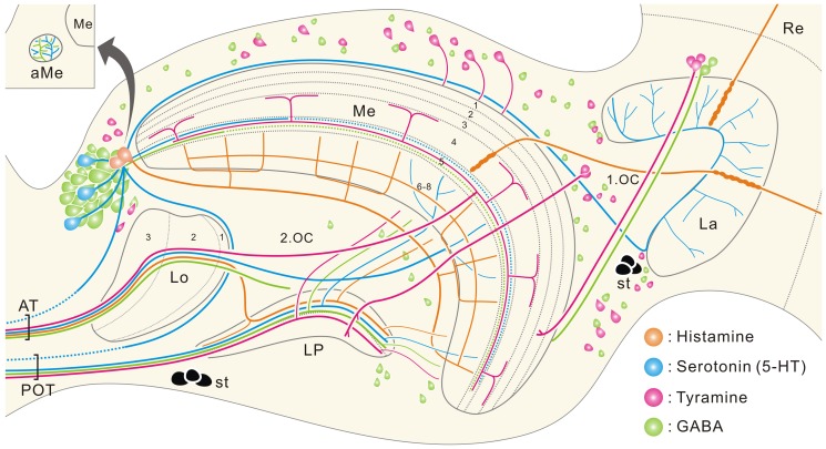
Figure 12. Schematic illustration depicting the distribution of immunolabeled cell bodies and major fiber trajectories.
Horizontal plane. Anti-histamine in orange, anti-5-HT in blue, anti-tyramine in magenta, and anti-GABA in green. In the medulla (Me), 5-HT-positive fibers in layers 1–4 and GABA-positive fibers in layers 1–4 and 6–8 are omitted for clarity. In the lobula (Lo) and lobula plate (LP), likewise, immunoreactive arborizations in respective layers are omitted. Solid lines indicate confirmed pathways, and dotted lines are putative pathways. 1.OC, first optic chiasma; 2.OC, second optic chiasma; aMe, accessory medulla; AT, anteriorly running optic tracts; La, lamina; POT, posterior optic tract; Re, retina; st, larval stemmata.

