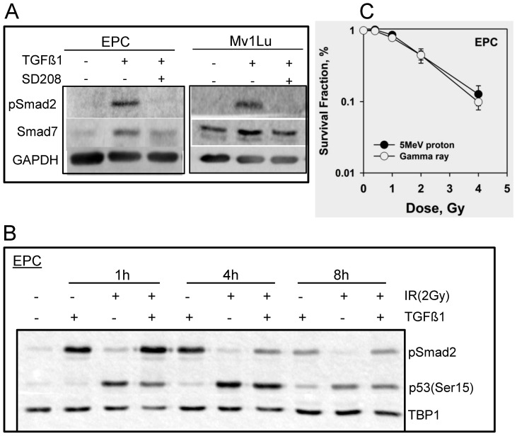
Figure 1. Cellular response to TGFβ1 and IR exposure in EPC and Mv1Lu cells.
A. Whole cell extracts were prepared 4 h after 2 ng/ml TGFβ1 treatment using M-PER kit. Phosphorylation of Smad2 and expression of Smad7 were detected using western blotting. TGFβR1 inhibitor- SD208 (or DMSO as control) was added one hour prior to the addition of TGFβ1. GAPDH was used as loading control. B. Nuclear extracts were isolated from EPC cells using NE-PER kit after 2 Gy of proton radiation at indicated times, in the presence or absence of TGFβ1. Shown are the western blots with phosphorylation of Smad2 and p53, and TBP1 as internal loading control. C. Clonogenic survival assay of EPC cells with different doses of protons and gamma rays as described in Materials and Methods.

