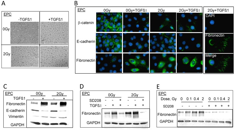
Figure 3. Protons enhance EMT induced by TGFβ1 in EPC cells.
A. Phase contrast images of morphology alterations after double treatment of TGFβ1 and 2 Gy of protons. B. Immunostaining of EMT markers, 3 days post 2 Gy of protons in the presence or absence of TGFβ1. Right panel shows the tracks of fibronectin staining in EPC cells after double treatment. C. Western blots show the change of EMT markers expression upon TGFβ1 and/or protons treatment. Whole cell extracts were prepared using RIPA buffer. D. Western blots show the inhibition of fibronectin expression when EPC cells were pretreated with TGFβR1 inhibitor-SD208 1 h before 2 Gy or 0 Gy of proton radiation. E. The inhibition effect of SD208 on protons treatment alone.

