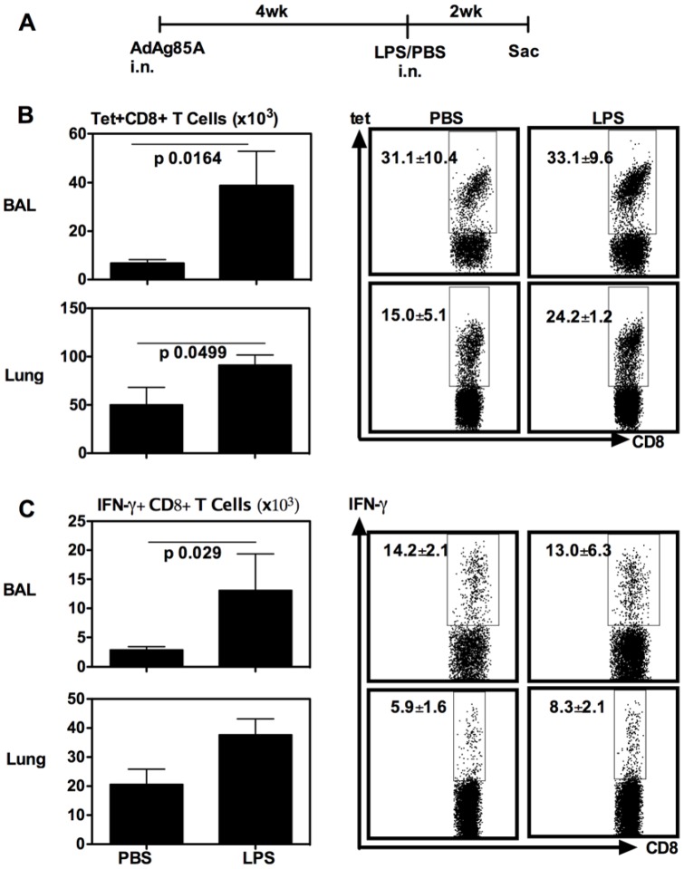
Figure 2. Respiratory LPS exposure increases vaccine-induced T cells in airway lumen (BAL) and lung interstitium at 14 days.
(A) Experimental schema; At 14 days post-LPS or -PBS, mice were sacrificed and BAL and lung cells were isolated, and Ag85A tetramer (Tet)-specific (B) and IFN-γ-secreting (C) CD8+ T cells in BAL or lung interstitium were determined by immunostaining, intracellular cytokine staining and FACS. Representative dotplots are shown with the average frequency ± SEM from three animals/group. The data in graphs are expressed as mean value ± SEM of three animals per group, representative of three independent experiments.

