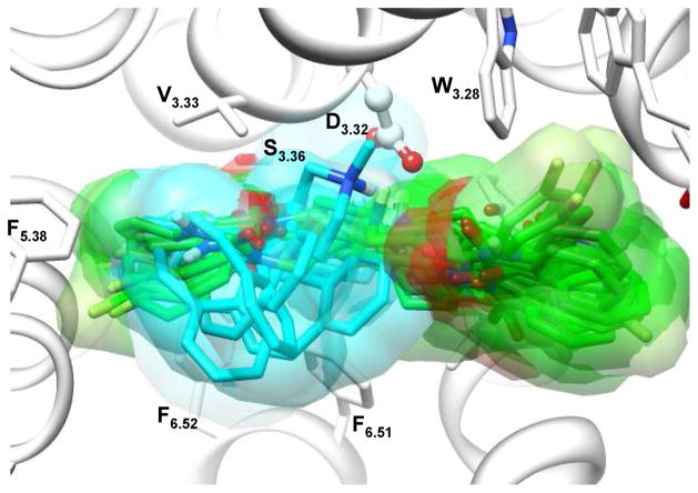
Figure 3.
Overlapped diverse representative docking poses of ketanserin and cyproheptadine in the binding-site of the initial homology model of 5-HT2A. Cyproheptadine (carbon atoms colored cyan) mainly binds in site 1, mainly bordered by TM3, 5 and 6, while ketanserin (colored green) adopts extended conformations that allow binding both sites.

