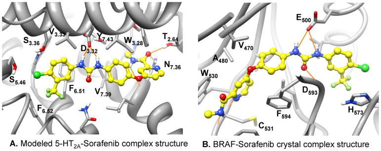
Figure 8.
The binding mode of sorafenib in the modeled 5-HT2A-sorafenib complex structure and the BRAF-sorafenib crystal complex structure. Carbon atoms of sorafenib are colored in yellow. The hydrogen bond interactions between the urea group of the sorafenib and the conserved D3.32 in 5-HT2A (A) or the catalytic residue E500 in BRAF (B) are illustrated with orange line. Molecular images were generated with UCSF Chimera.

