
Figure 1.
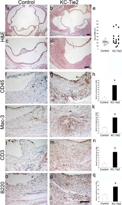
Aortic roots develop spontaneous vascular inflammation by 1 year of age in KC-Tie2 and not control animals. Aortic roots from control (a, c) and KC-Tie2 (b, d) animals stained with H&E. Quantitation of aortic root vessel wall area (e) demonstrates significant increases in area of KC-Tie2 mice (n=19) compared to control littermates (n=18). Immunohistological staining and quantitative analyses of positively stained CD45+ (f–h), Mac-3+ (i–k), CD3+ (l–n) and B220+ cells (o–q) (% positively stained area) within the aortic roots of representative control (f, l, i, o; n = 5) and KC-Tie2 mice with vascular inflammation (g, j, m, p; n=5) reveals significant increases in inflammatory cell infiltrates. * P <0.05 vs control animals. Scale bar = 100 μm.