
Figure 2.
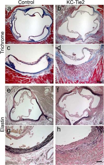
Anatomical characterization of aortic arch lesions. Aortic roots from control (a, c, e, g) and KC-Tie2 (b, d, f, h) animals stained with Trichrome (a–d) or Verhoeff–van Gieson elastin (e–h) reveals decreased expression of collagen in Trichrome stained aortic arch tissue from KC-Tie2 mice and increases in the numbers of elastin breaks in Verhoeff–van Gieson elastin stained aortic root tissues. Scale bar = 100 μm.