
Figure 3.
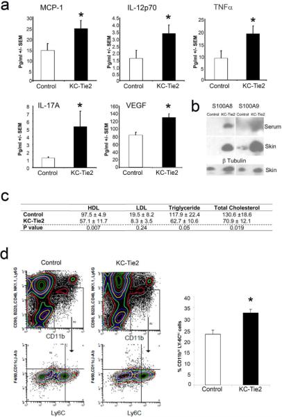
Aortic root vascular inflammation develops in the presence of elevated systemic inflammation and monocytosis and independent of lower lipid levels. Proinflammatory cytokines are significantly elevated in KC-Tie2 mouse sera, including levels of MCP-1, IL-12p70, TNFα, IL-17A and VEGF (a; mean ± SEM; n=5–11 per group) and S100A8/A9 expression is increased in skin and serum of KC-Tie2 animals (b). KC-Tie2 animals have significantly less total cholesterol, triglycerides and HDL compared to control mice (c; mean ± SEM; n=8 per group; p values indicated for each lipid in table). 4-colour flow cytometry reveals significant increases in splenic proatherogenic monocytes (CD90loB220loCD49bloNK1.1loLy6GloCD11cloIAbloF4/80loCD11bhiLy-6Chi). Representative flow cytometry and quantification of CD11b+F4/80loLy-6Chi cells (d; mean ± SEM; n =4 spleens per group).* P <0.05 vs. control animals.