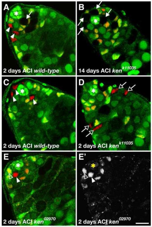
Figure 2. ken is required cell-autonomously for CySC but not GSC maintenance.
(A-E) Confocal sections through the testis apex. Hubs denoted by asterisks. (A) Wild-type GSC clone (arrow) adjacent to the hub identified by absence of GFP (green) and Tj (red) at 2 days after clone induction (ACI). Wild-type CySC clones (arrowheads) identified by absence of GFP (green) and presence of Tj (red) are visible in this testis as well. (B) kenk11035 mutant GSC clones (arrows) are still present 14 days ACI and producing normal differentiating spermatogonia whereas there are virtually no remaining kenk11035 CySC clones by this time point. (C) Wild-type CySC clones (arrowheads) surrounding the hub at 2 days after clone induction (ACI). (D) Differentiating kenk11035 mutant cyst cell clones (open arrows) identified by absence of GFP (green), presence of Tj (red), and distance from the hub. There are no kenk11035 CySCs surrounding the hub indicating that these cyst cells originated from CySC clones generated 2 days prior. (E) ken02970 mutant CySC clone (arrowhead) 2 days ACI identified by the absence of GFP (green) and presence of ZFH1 (red) has similar ZFH1 levels relative to neighboring wild-type CySCs (one indicated, open arrowhead). (E’) ZFH1 channel alone. Scale bar, 10 m.

