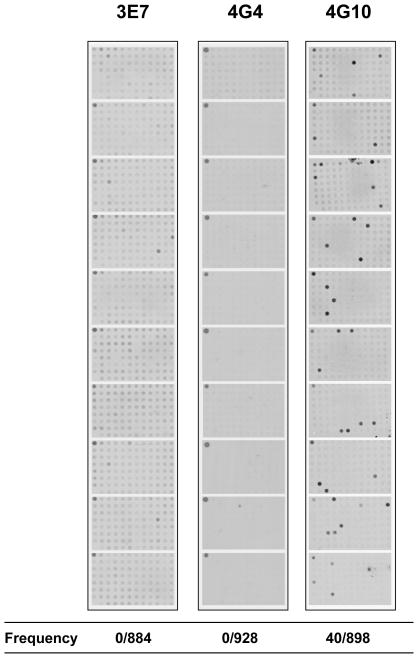
Figure 4.
B cell clones frequencies in the blood. Frequency of the 3 polyreactive clones was assessed in the blood by hybridization of plasmid cloned PCR amplified VH segments using dot blot assays as described in Figure S3. Bacterial colonies grown in 96 well microplates were transferred onto membranes and screened with the corresponding clonotypic probes (Figure S2). Ten membranes were screened for each clone. The upper left corner dot in each plate contained a control plasmid. Each positive dot was further analyzed to verify the presence of the unique clonal CDR3 sequence. All weakly positive dots observed for 3E7 were false positive. Each membrane was also screened for the presence of a VH sequence using a consensual JH probe. The ratio of positive clonotypic sequence on total VH sequence is reported for each clone.

