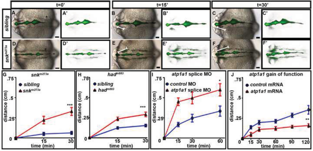
Fig. 2. Atp1a1 regulates neuroepithelial permeability.
(A–F) Dye retention assay in sibling embryos (A–C) vs. snkto273a mutants (D–F). Brightfield dorsal views of embryos ventricle injected with a 70 kDa FITC Dextran (A–F) and corresponding dye only images (A’–F’) over time. The distance the dye front moves measured at each time point indicated by white line. (G–J) Quantification of permeability in snkto273a mutants (G), hadm883 (H), atp1a1 splice site morphants (I), and atp1a1 gain-of-function embryos (J) compared to control injected. Average taken from 3–6 independent experiments and represented by mean +/− SEM. ***= p<0.0001, **= p<0.005, *=p<0.05 compared to control. All images taken at 22–24 hpf with anterior to left. Asterisk = ear. Scale bars = 50µm.

