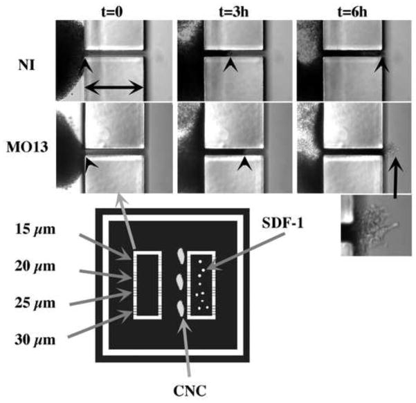
Figure 7. CNC cells can migrate through 20 μm openings.
Migration of CNC through small openings. Photographs of explants (10X) taken from time-lapse movies are represented. The width of the obstacle is 200 μm (horizontal arrows) while the opening is 20 μm. Frames from non-injected control (NI) or morphant (MO13) CNC are presented at 0, 3 and 6 hours of migration. The arrowhead points to the leading edge of cells migrating in the tunnel. The mask used to obtain the substrates is represented below. The white sections represent the walls of the chambers (200 μm high). Each of the two rectangular chambers has four opening for each of their long side ranging from 15 to 30 μm). The center of the rectangles are filled with SDF-1-coated beads and sealed by covering it with a small coverslip fixed with vacuum grease. Explants are placed with their ventral (leading) edge facing each opening.

