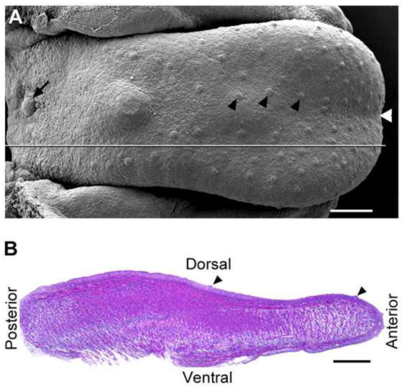
Fig. 1.

A: Scanning electron microscopy image illustrates the dorsal view of an E15.5 embryonic tongue and papilla types. Black arrowheads point to fungiform papillae on the anterior oral tongue; black arrow points to the single circumvallate papilla in the back. White arrowhead at the tip points to the median furrow. The straight line marks the orientation for sectioning in the sagittal plane. B: H&E stained sagittal section of an E15.5 tongue to illustrate the orientation for all images of tongue sections. Black arrowheads point to fungiform papillae. Scale bars: 250 μm.
