
Fig. 7.
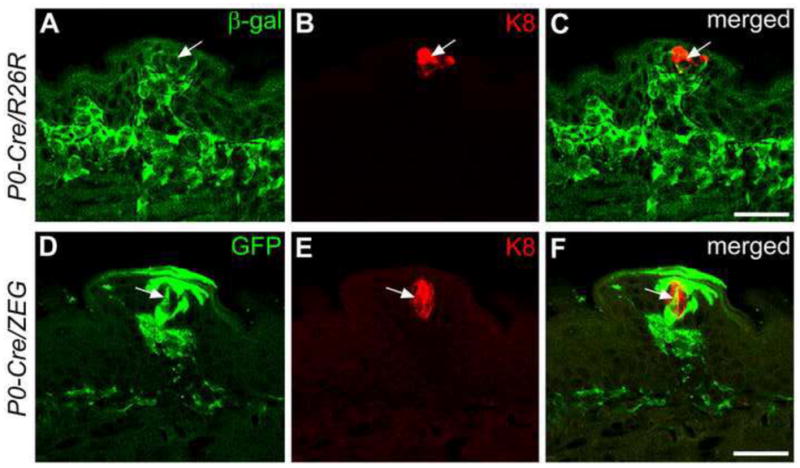
Single plane confocal images of fungiform papillae and taste buds with double labeling of β-gal (A) or GFP (D) (green) and K8 (B, E, red) immunoreactions at P1–5 in P0-Cre driven R26R (A–C) or ZEG (D–F) mouse tongue. β-gal and GFP positive cells are observed within taste buds and some of the cells (white arrows) are also labeled with K8. Scale bars: 25 μm.