
Fig. 8.
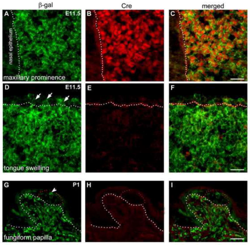
Maxillary prominence (A–C) and tongue swelling (D–F) at E11.5 and fungiform papillae at P1 (G–I) in P0-Cre/R26R mouse line. Sections were double labeled with antibodies against β-gal (green) and Cre (red). Anterior tip of the upper jaw and tongue are toward to the right. White dotted lines demarcate the border between epithelium and mesenchyme. Red dots outline early taste buds in fungiform papilla at P1 (G–I). Short arrows point to β-gal labeled epithelial cells (D) and arrowhead (G) points to labeled cells in early taste buds. Scale bars: 25 μm.