
Fig. 9.
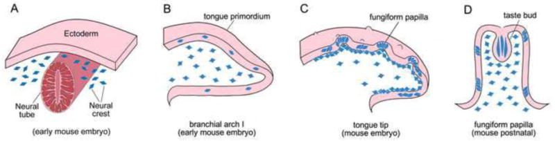
Diagram to illustrate distribution of NC cells and NC-derived cells in developing tongue. A: Cross section of an early stage embryo at a cranial level. The delaminated, migratory NC cells (blue) are located at both sides lateral to neural tube (red), under the ectodermal sheet (pink). B: Sagittal section of branchial arch I (tongue primordium) before tongue emerges. The anterior tip is toward to the right and dorsal surface up (also applies to C and D). Migrated NC cells (blue) are scattered in the epithelium and broadly distributed in the mesenchyme. C: Sagittal section of anterior region of oral tongue with developing fungiform papillae on the dorsal surface. NC derived cells (blue) are clustered in tongue epithelium within and between papillae. In tongue mesenchyme, NC derived cells are more restricted under the epithelium. D: A fungiform papilla with single taste bud at the apex. NC derived cells are within taste buds and in the surrounding papilla epithelium. Also, NC derived cells are densely distributed in the mesenchymal core of papillae. We propose that NC cells (A, blue cells) migrate into the epithelium and mesenchyme of tongue primordium (B) at early embryonic stage. The NC cells in tongue epithelium acquire epithelial phenotype and undergo cell proliferation and differentiation to become clusters within and between papillae (C). A population of NC-derived cells is within early taste buds (D). In tongue mesenchyme, NC-derived cells are progressively restricted to connective tissues under tongue epithelium and densely distributed in the mesenchymal core of taste papillae (C, D).