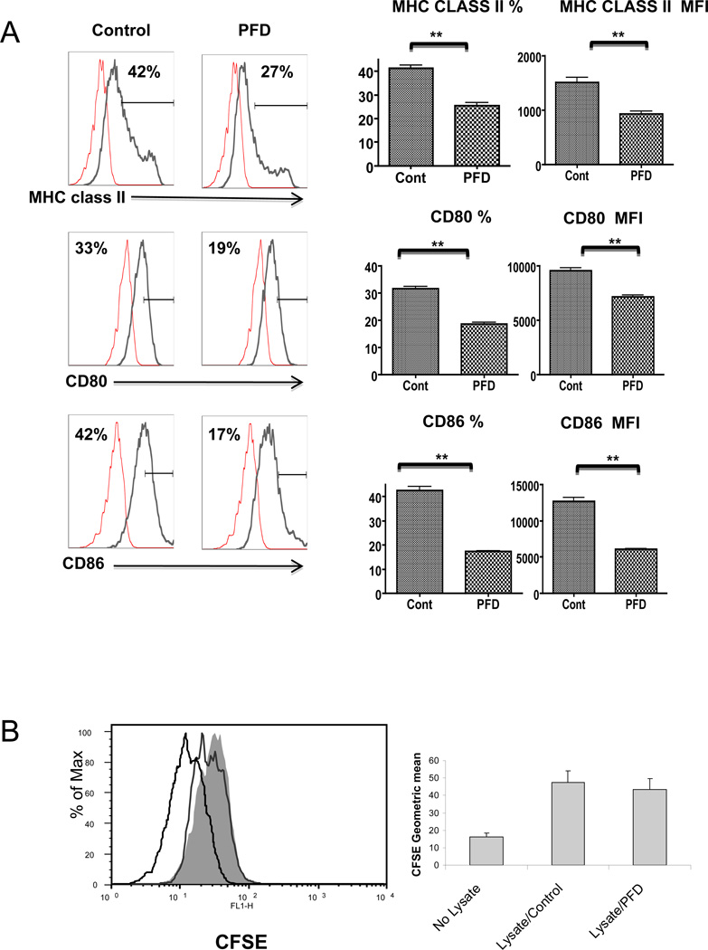
Figure 3.
A. PFD inhibits DC maturation and activation in vitro;
DCs were incubated with or without 2 mM PFD for 72 hours, stimulated with 1µg/ml LPS, and evaluated 24 h later based on MHC class II, CD80, and CD86 expression using flow cytometry. The PFD treated DCs express significantly less MHC class II and the co-stimulatory molecules CD80 and CD86 as compared to control/untreated cells. Presented are representative flow cytometry histograms showing mean fluorescence intensity (MFI) of the staining for each of the antibodies along with isotype control and graphs for percentage of MHC class II, CD80 and CD86 positive cells and MFI of each based on the mean +/− SEM of 4 independent experiments.
B. PFD does not inhibit DCs alloantigen uptake;
C57BL/6 BM derived DCs were incubated with or without 2mM PFD for 72 h. DCs were then exposed to CFSE pre-labeled BALB/c splenocyte lysates at 1:5 ratio overnight. DC ability to uptake was assessed by flow cytometry for CFSE labeling within DCs. PFD treatment had no significant effect on DCs ability to uptake alloantigen. A representative histogram is shown illustrating negative control (no CFSE), CFSE staining with (unshaded) and without (shaded) PFD treatment along with a graph illustrating the MFI mean +/− SE for the ability of C57BL/6 DC to endocytose CFSE pre-labeled BALB/c splenocyte lysate.

