
Figure 3. Mutational analysis of CTNNB1 and WT1 in Kenyan WT.
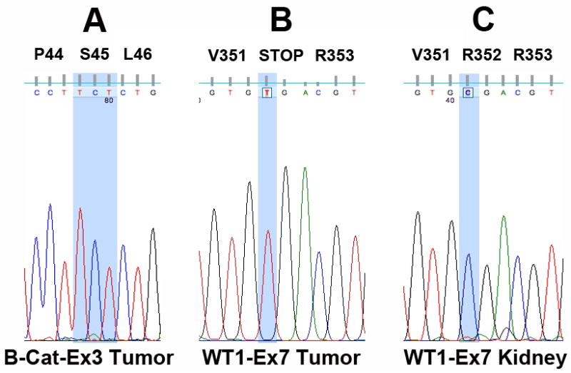
(A) Despite high levels of β-catenin nuclear expression, mutational analysis revealed wild-type β-catenin in all cases at exons 3, 7, and 8. The wild-type sequence coding Serine 45 (S45), a critical phosphorylation residue in exon 3, is highlighted. (B) A single mutation (p.Arg352X; c.1244 C>T) predicted to alter the function of WT1 was detected in specimen 10. The homozygous C to T mutation responsible for conversion of Arginine to a stop codon is highlighted. (C) The wild-type sequence corresponding to (B) was detected in adjacent areas of normal kidney.