
Figure 1. Cell death in primary cultured cortical neurons from male and female rat brains.
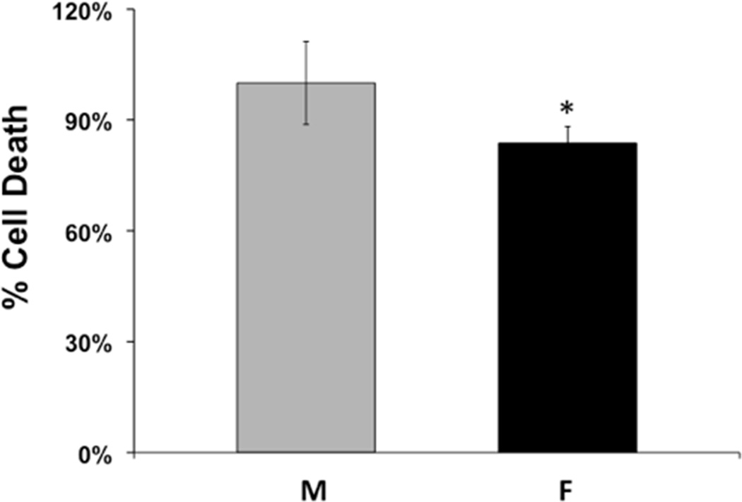
Cortical neurons were cultured from embryonic day 18 male (M) and female (F) rat embryos and were subjected to OGD on day 10 in vitro. Cell death was measured by LDH release in neurons from male and female rats 24 hrs after reoxygenation. The graph depicts average cell death in neurons from male and female embryos expressed relative to the mean of cell death in males. (n=9, *p=0.0019).