
Figure 4. Western blot analysis of soluble epoxide hydrolase in cortical neurons from males and females.
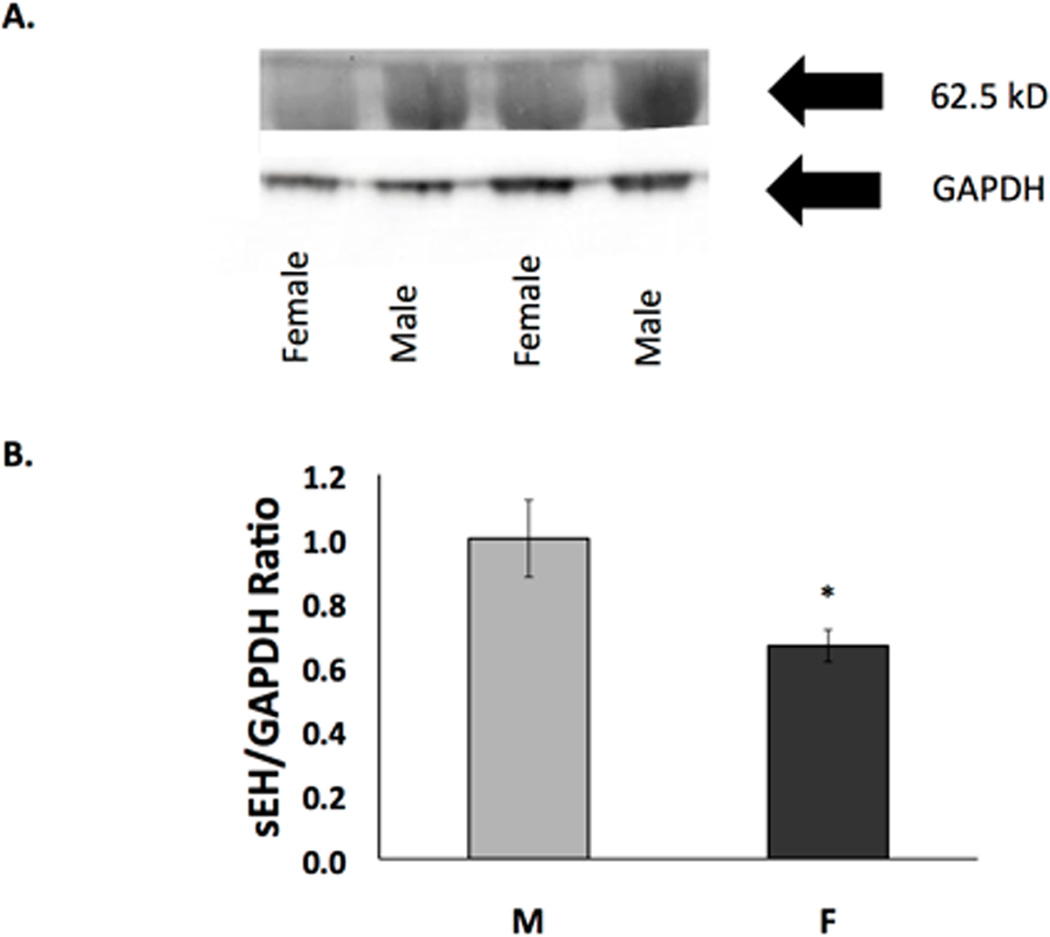
Western blot of protein extracts of neurons from males and females harvested after 10 days in vitro. sEH expression in cultured cortical neurons from males is significantly higher than in neurons from females. (A) Representative image of Western blot. Top panel is probed with anti-sEH antibody and bottom panel is probed with anti-GAPDH. (B) Quantification of Western blots of sEH normalized against GAPDH. Cells from males and females were paired within each experiment (n=4, *p=0.004)