
Figure 6. 14,15-EET was present in cells from males and females at baseline and after OGD.
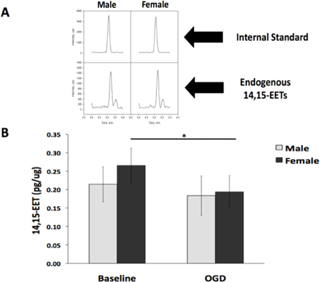
The concentration of EETs in neurons from males and females was evaluated using liquid chromatography-tandem mass spectrometry (LC-MS/MS) in untreated (baseline) cells and cells subjected to 2 hours of OGD followed by 24 hours reoxygenation. (A) Representative 14,15-EET peaks obtained via LC-MS/MS in both male (M) and female (F) cells. The top panel represents the d8 14,15-EET internal standard. The bottom panel shows the endogenous 14, 15-EET peaks from the cells. The left two are from male cells and the right two are from female cells. The SRM transitions monitored were m/z 327.2 to 182.2 for the internal standard and the m/z for the 14,15 EET is 319.2 to 175. (B) The concentration of total 14,15-EET is not significantly different in cells from males and females at baseline (n=5, p=0.07). After OGD, 14,15-EET significantly decreased in cells from females, but not males. * Indicates a significant decrease in 14,15-EET after OGD in cells from females (n=5, *p=0.015).