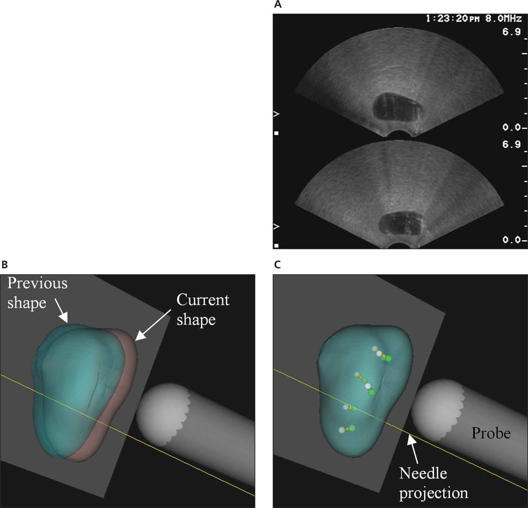
Figure 1.
Comparison between 2D and 3D displays of biopsy procedures. A, Screen capture of sonograms of a prostate phantom. Only 2 views, transverse (top) and sagittal (bottom), are visible. B and C, Screen captures from the Eigen Artemis system showing a subsequent biopsy procedure. B, Unregistered previous (green) and current (pink) volumes. C, Registered volumes with previous (white) and current (green) core locations inside.

