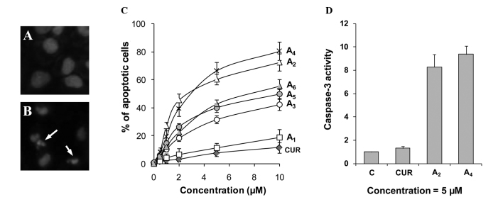
Figure 2.
Effects of curcumin analogues on apoptosis. PC-3 cells were seeded at a density of 0.2×10⁵ cells/ml of medium in 35-mm tissue culture dishes (2 ml/dish) and incubated for 24 h. The cells were then treated with various concentrations (0.5–10 μM) of the different compounds for 72 h. (A and B) Representative micrographs of propidium iodide-stained controls and A4 (5 μM)-treated PC-3 cells. Arrows indicate apoptotic cells. (C) Percentage of apoptotic cells as determined by morphological assessment in PC-3 cells treated with the various compounds. (D) Caspase-3 activities in PC-3 cells treated with curcumin, A2 and A4. Each value is the mean ± SD from three experiments. C, control; CUR, curcumin.

