
Figure 1.
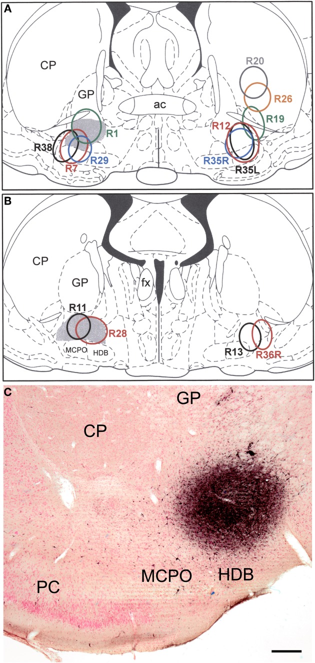
(A and B) Drawings showing the FG injection sites into the BF (and overlying striatum) plotted at the Bregma −0.3 (A) and Bregma −0.8 (B) levels of the atlas by Paxinos and Watson (1997). Gray shading indicates the SI/VP portion of the BF, the main source of the cholinergic innervation of the amygdala. Injections in rats whose brains were processed for triple-labeling immunofluorescence (FG/SOM/NPY and FG/SOM/CB) are depicted on the left. Injections in rats whose brains were processed for double-labeling immunofluorescence (FG/SOM, FG/PV, and FG/CR) are depicted on the right. The letters “L” (left) and “R” (right) in three of the case numbers (R35L, R35R, R36R) indicate whether the injection site in these three bilaterally injected rats was on the left or right side. (C) Photomicrograph of the injection site in case R28. This section was counterstained with pyronin Y (a pink Nissl stain). Left is lateral. Abbreviations: ac, anterior commissure; CP, caudate-putamen; fx, fornix; GP, globus pallidus; HDB, horizontal limb of the nucleus of the diagonal band; MCPO, magnocellular preoptic nucleus; PC, piriform cortex. Scale bar in C = 250 μ m.