
Figure 10.
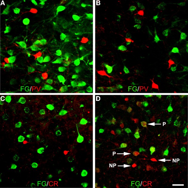
Photomicrographs of immunofluorescence preparations dual-labeled for FG/PV or FG/CR. (A) Dual-localization of FG (green) and PV (red) in the anterior basolateral nucleus. Note lack of colocalization. (B) Dual-localization of FG (green) and PV (red) in the posterior basomedial nucleus. Note lack of colocalization. (C) Dual-localization of FG (green) and CR (red) in the anterior basolateral nucleus. Note lack of colocalization. (D) Dual-localization of FG (green) and CR (red) in the posterior basomedial nucleus. Note lack of colocalization in small non-pyramidal neurons exhibiting robust CR-ir (NP), but existence of colocalization in larger pyramidal neurons exhibiting light CR-ir (P). Scale bar in D= 40 μ m (also applies to A, B, and C).