
Figure 9.
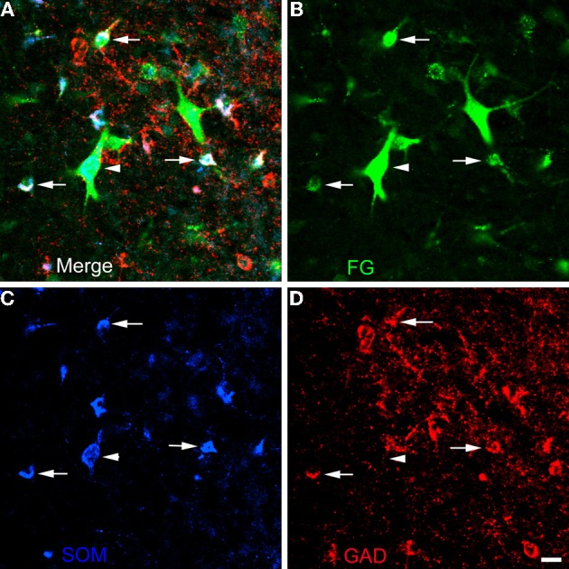
Photomicrographs of neurons in the external capsule laterally adjacent to the ventral basolateral nucleus at bregma level-2.8 in a section triple-labeled for FG (green), SOM (blue), and GAD (red). (A) Merged image visualizing all three channels. Arrows indicate 3 of 6 neurons in this field triple-labeled for FG/SOM/GAD+ (white). Arrowhead indicates a GAD-negative FG/SOM+ neuron (blue-green). The locations of the same neurons are indicated by identical arrows and arrowheads in B, C, and D. (B) Green channel image showing FG+ neurons. (C) Blue channel image showing SOM+ neurons. (D) Red channel image showing GAD+ neurons. Scale bar in D= 20 μ m (also applies to A, B, and C).