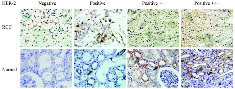
Figure 1.
Immunohistochemical staining of RCC and normal kidney tissues. HER2 expression in either RCC or normal renal tissues varies from negative to moderately or intensively positive. It should be noted that HER2 was mainly expressed in the renal tubule in normal tissues, as indicated by the arrow. Original magnification, ×400. RCC, renal cell carcinoma.

