
Figure 3.
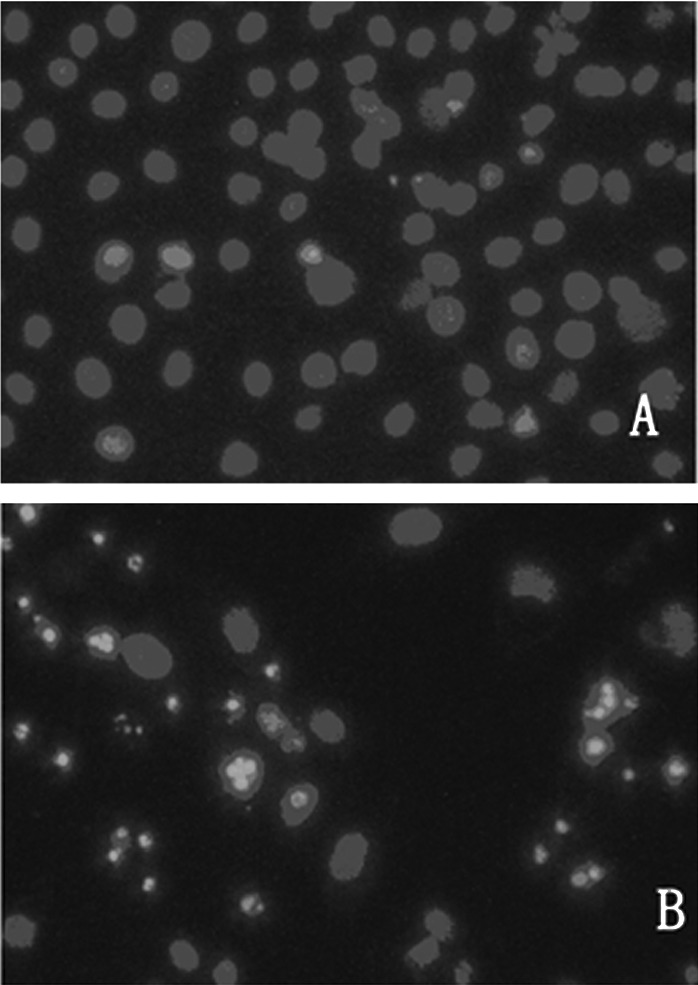
Fluorescence photomicrograph of MG-63 cells stained with Hoechst 33258. MG-63 cells were exposed to 50 μM of zoledronic acid for 72 h, harvested and cytospun onto glass slides for fixing. The preparations were stained with Hoechst 33258 and examined under fluorescence microscope. (A) Control; the nuclei were stained homogeneously and were less bright. (B) Treated cells; chromatin condensation occurred and apoptotic bodies formed. Magnification ×200.