
Figure 4.
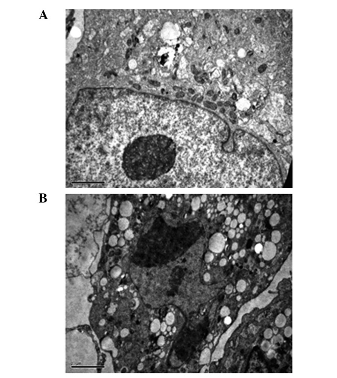
Electron micrographs of stained MG-63 cells. MG-63 cells were fixed in 2.5% glutaraldehyde, postfixed in 2% osmium tetroxide and embedded in Luveak-812. Ultrathin sections were stained with lead citrate and uranyl acetate and examined using a JEM-1230 electron microscope. Electron micrographs of (A) untreated cells and (B) cells treated with 50 μM ZOL for 72 h. (Scale bar, 10 μm).