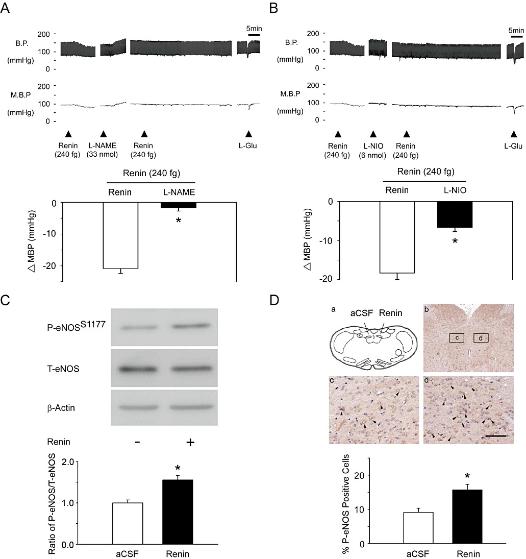
Figure 2.

Microinjection of renin induces eNOS-Ser1177 phosphorylation in the NTS. (A) Representative tracing demonstrates that the depressor effect of renin was significantly attenuated by a non-selective NOS inhibitor, L-NAME (33 nmol). Summary data (means ± SEM, n= 6) are shown in the graph. *P < 0.05 significantly different from the renin group. (B) Representative tracing demonstrates that the depressor effect of renin was significantly attenuated by the specific eNOS inhibitor, L-NIO (6 nmol). Summary data (means ± SEM, n= 6) are shown in the graph. *P < 0.05, significantly different from the renin group. (C) The quantitative immunoblotting analysis demonstrates that renin treatment increased the level of P-eNOS-Ser1177 protein in the NTS. Densitometric analysis of P-eNOS-Ser1177 protein levels (means ± SEM, n= 6) after administration of aCSF or renin. *P < 0.05, significantly different from the aCSF group. (D) Immunohistochemical staining of the brainstem for P-eNOS-Ser1177 showed that injection of renin into the NTS induced P-eNOS-Ser1177 (c vs. d). Arrows indicate P-eNOS-Ser1177-positive cells. The scale bar represents 200 µm. Summary data (means ± SEM, n= 6) are shown in the graph. The percentage of P-eNOS-Ser1177-positive cells was determined by counting P-eNOS-Ser1177-expressing cells in each hemisphere of the NTS at 200 × magnification. These counts were divided by all of the cells in the same paraffin section. *P < 0.05, significantly different from the a CSF group.
