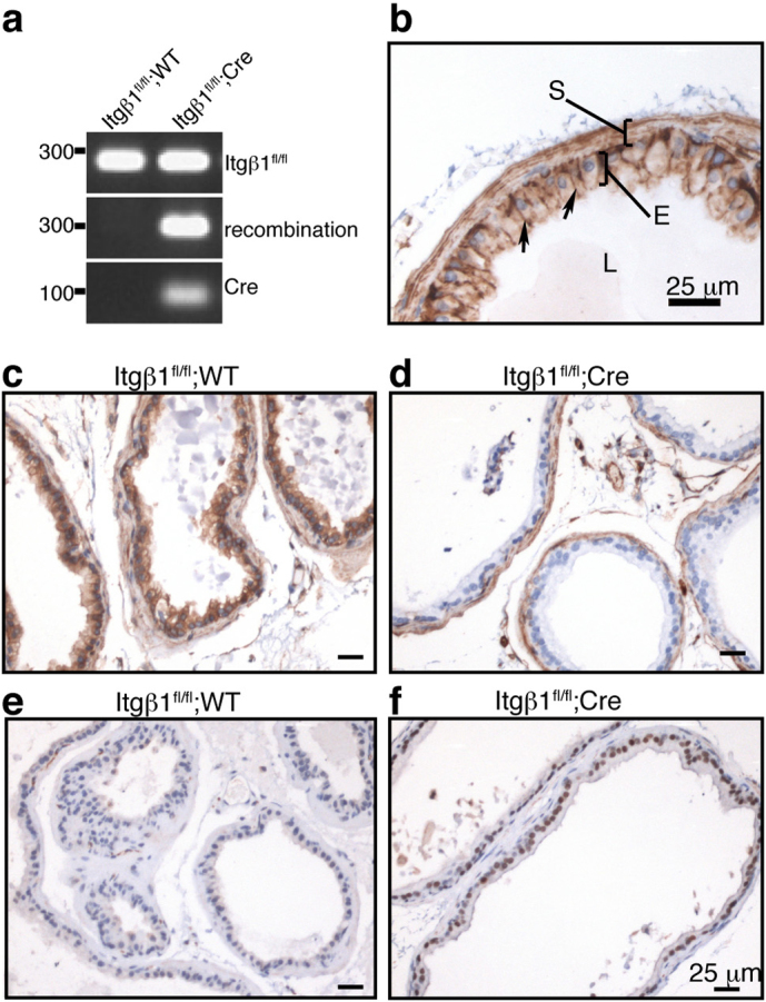
Figure 1. Specific deletion of β1 integrin in prostate epithelium.

a) PCR analysis of Cre-mediated recombination in prostate glands of Itgβ1fl/fl;WT and Itgβ1fl/fl;Cre animals at 26 weeks of age. The 280 bp product labelled Itgβ1fl/fl indicates the presence of the floxed allele. Also shown are the 300 bp amplicon resulting from recombination of the floxed allele, and the 100 bp product demonstrating the presence of the Cre transgene. b) β1 integrin immuno-staining in the dorsal prostate of a 20 wk WT mouse. β1 integrin is present predominantly on the baso-lateral surfaces of the luminal epithelial (E) cells (arrows), but is also present at the cell membrane of stromal cells (S). c & d) β1 integrin staining of the ventral prostates of 26 wk old Itgβ1fl/fl;WT (c) and Itgβ1fl/fl;Cre (d) animals, demonstrating almost complete ablation of β1 integrin within the epithelium (d), but intact stromal staining. e) Cre staining of prostates of 26 wk old Itgβ1fl/fl;WT (e) and Itgβ1fl/fl;Cre (f) animals. Scale bars in b, c and d, 25 μm.
